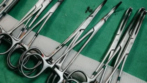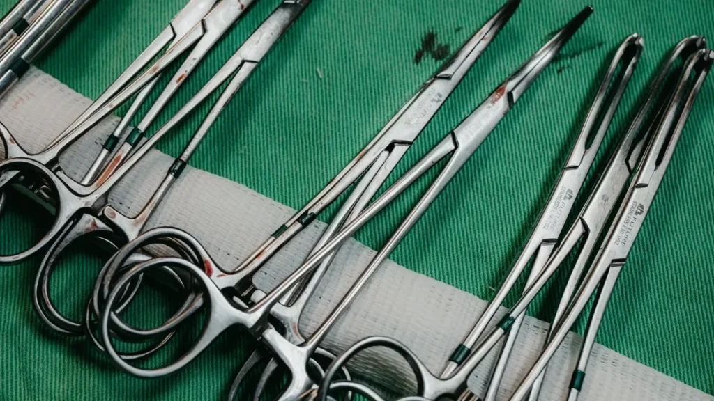
Here are 10 essential instruments used in laparoscopic surgery:
- Laparoscope
- Camera system
- Insufflator
- Trocar
- Graspers
- Dissector
- Laparoscopic scissors
- Energy devices
- Needle driver
- Surgical mesh
Learning about these essential instruments helps you make careful moves. You can handle tissues the right way. This also keeps patients safe by causing less harm.
Key Takeaways
- Knowing important tools like the laparoscope and trocars is very important for safe surgery. Each tool has a special job. These tools help lower harm to patients and make surgery more accurate.
- Using the correct tools, like graspers and energy devices, helps surgeons do careful work well. This means patients get better faster and have fewer problems after surgery.
- Practicing often and knowing these tools well makes surgeons more confident and skilled. Being good with laparoscopic instruments helps patients stay safe and have better results during surgery.
Essential Instruments Overview
When you begin learning laparoscopic surgery, you must know the main instruments. These tools help you look inside the body. They also help you hold and move tissues. Each tool does something important. You need all of them to do surgery safely.
Instrument List
Always use the right tools. This makes your job easier and safer.
Here is why these tools are important:
- They help you see inside the body better.
- They let you move and cut tissues gently.
- They lower the chance of hurting the patient.
- They make small cuts, so patients get better faster.
You use these tools in almost every laparoscopic surgery. You should learn how each one works. You also need to practice using them. When you know these tools well, you can do your best work. You can also keep your patients safe.
If you want to be good at laparoscopic surgery, you must learn about these tools. You will use them every time you do surgery. Knowing what they are and how they work will help you feel sure of yourself.
Laparoscope
Description
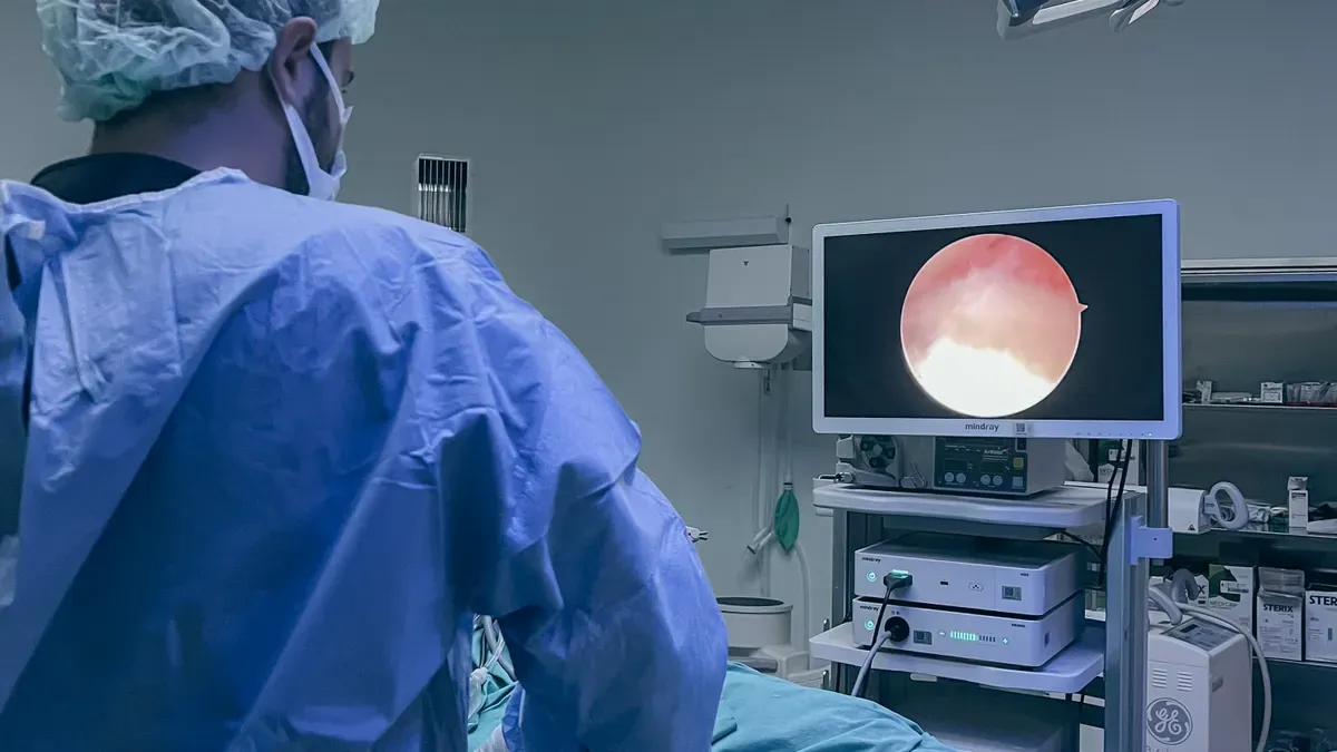
A laparoscope helps doctors see inside the body. It is a thin tube with a camera and light. Doctors put it in through a small cut. The camera shows pictures on a screen. This lets doctors watch what they do. There are different types for different surgeries.
| Type of Laparoscope | Description |
|---|---|
| Laparoscope | Used for abdominal cavity surgeries. |
| Thoracoscope | Used for chest cavity surgeries. |
| Arthroscope | Used for joint surgeries. |
Function
Doctors use the laparoscope to guide their actions. It helps them see organs and tissues. They do not need to make big cuts. Doctors can find problems and move tools. They can do careful work. The laparoscope is important for safe surgery.
Features
A laparoscope has some key features:
- High-definition camera shows clear pictures
- Bright light helps doctors see better
- Long, slim shape makes it easy to use
- Magnifies the view for more detail
Tip: The laparoscope lets doctors see more than open surgery. They can make smaller moves and be safer.
Here are some good things about using a laparoscope:
| Advantage | Description |
|---|---|
| Enhanced Precision and Visualization | Laparoscopy gives a bigger view, so doctors see better and work carefully. |
| Faster Recovery Times | Patients get better faster because there is less harm and blood loss. |
| Reduced Surgical Trauma and Blood Loss | Small cuts mean less harm and fewer blood transfusions, so recovery is quicker. |
| Lower Risk of Post-Surgical Complications | Laparoscopic surgery lowers the chance of problems after surgery. |
| Improved Cosmetic Outcomes | Small cuts leave less scars than regular surgery. |
Camera System
Description
The camera system helps you see inside the body. It connects to the laparoscope and sends pictures to a screen. This system has many parts that work together. You will find the imaging system, light source, light cable, camera, and monitor in every setup. Each part helps you see clearly and makes your job easier.
| Component | Description |
|---|---|
| Imaging System | Takes pictures and sends them to the monitor. |
| Laparoscope | Makes the view bigger inside the body, comes in different sizes and angles. |
| Light Source | Gives bright light, often using halogen or xenon bulbs. |
| Light Cable | Brings light from the source to the surgery area using optical fibers. |
| Camera | Sends pictures from the laparoscope to the monitor. |
| Monitor | Shows live pictures for you to watch during surgery. |
Function
The camera system lets you see organs and tissues in detail. You can watch your actions on the screen and make careful choices. High-resolution cameras help you spot tiny blood vessels and nerves. You avoid mistakes because you see everything clearly. The camera system is one of the Essential Instruments that keeps laparoscopic surgery safe and accurate.
Tip: Always check the camera system before surgery. Clear pictures help you work faster and safer.
Features
Modern camera systems give you sharp and colorful pictures. Some systems show 3D views, which help you see how deep things are. You can switch between close-up and wide views so you do not miss anything. These features help you find problems early and stop injuries.
- High-resolution pictures help you make accurate moves.
- 4K and 3D technology help you see depth, important for hard tasks.
- Dual-view lets you see close and wide, making surgery safer.
You depend on these features to do careful work and keep patients safe.
Insufflator
Description
An insufflator fills the belly with carbon dioxide gas. This happens during laparoscopic surgery. The gas makes space inside the body. This helps you see organs and move tools. The insufflator connects to a tube. The tube goes through a small cut. Gas flows in at a set speed. You can change the settings for each patient. The device has a filter. The filter keeps the gas clean and free from germs.
Function
The insufflator makes a safe space to work. It gently pushes the belly wall away from organs. This gives you a clear view and more room. The filter blocks bacteria, viruses, and tiny bits. This lowers the chance of infection. It keeps the area clean. Using this device helps protect your patient. The insufflator is one of the Essential Instruments for safe surgery.
Tip: Always check the filter before you start surgery. Clean gas helps keep your patient safe.
Features
You can set the pressure and flow on the insufflator. Most devices let you pick the best settings. Lower pressure is better for patients. Here is a table with the suggested pressure settings:
| Pressure Setting | Description | Impact on Patient Outcomes |
|---|---|---|
| 15 mmHg or more | Used in the past for enough space | Lets CO2 out without losing space |
| 10 mmHg or less | Better for patient outcomes | Good for upper GI, gynecology, and colorectal surgery |
Modern insufflators have alarms and safety tools. You get alerts if pressure is too high or low. Some show numbers on a screen. You can watch everything easily. These features help you work safely and keep your patient comfortable.
Trocar
Description
A trocar helps doctors make a way into the body. It looks like a tube that can be sharp or blunt. Doctors push it through the skin and muscle. This lets them reach inside the body. Trocars come in many shapes and sizes. Some have blades, and some have cone tips. You can pick metal trocars that you use again or plastic ones you throw away. Optical access trocars let you see the tip as you push it in. Radially expanding access trocars stretch the opening softly. This lowers harm to the tissue.
- Types of trocars you might use:
- Metal trocars you can use again
- Plastic trocars you throw away
- Blunt trocars with cone tips
- Sharp trocars with special tips
- Optical access trocars for seeing the tip
- Radially expanding trocars for gentle entry
Function
Doctors use trocars to start most surgeries with small cuts. Trocars help put other tools inside, like graspers and cameras. You use trocars for many kinds of surgery. These include surgery on the appendix, gallbladder, intestines, liver, pancreas, and female organs. Trocars also work for chest surgery, draining fluid, and hormone pellet therapy. Animal doctors use trocars for animal surgeries and taking out fluid.
Tip: Always check which trocar you need before surgery. Picking the right one helps you avoid injuries and makes your job easier.
Features
How a trocar is made changes how safe and easy it is to use. You must pick the best one for each patient and surgery. Some trocars cut tissue fast, while others stretch the opening gently. The table below shows how trocar types are different:
| Trocar Type | Advantages | Disadvantages |
|---|---|---|
| Cutting Trocar | Goes in easily | More chance of hurting blood vessels or organs |
| Dilating Trocar | Less risk of injury | Needs more force, which can cause harm |
| Hybrid Trocar | Has both cutting and dilating features | May still need a lot of force to go in |
You should always use trocars carefully. Pick the right size and tip to lower the chance of hurting organs or getting a hernia. Optical access trocars help you see what you are doing, which keeps you safer. Radially expanding trocars stretch tissue instead of cutting, which helps stop injuries.
Graspers
Description
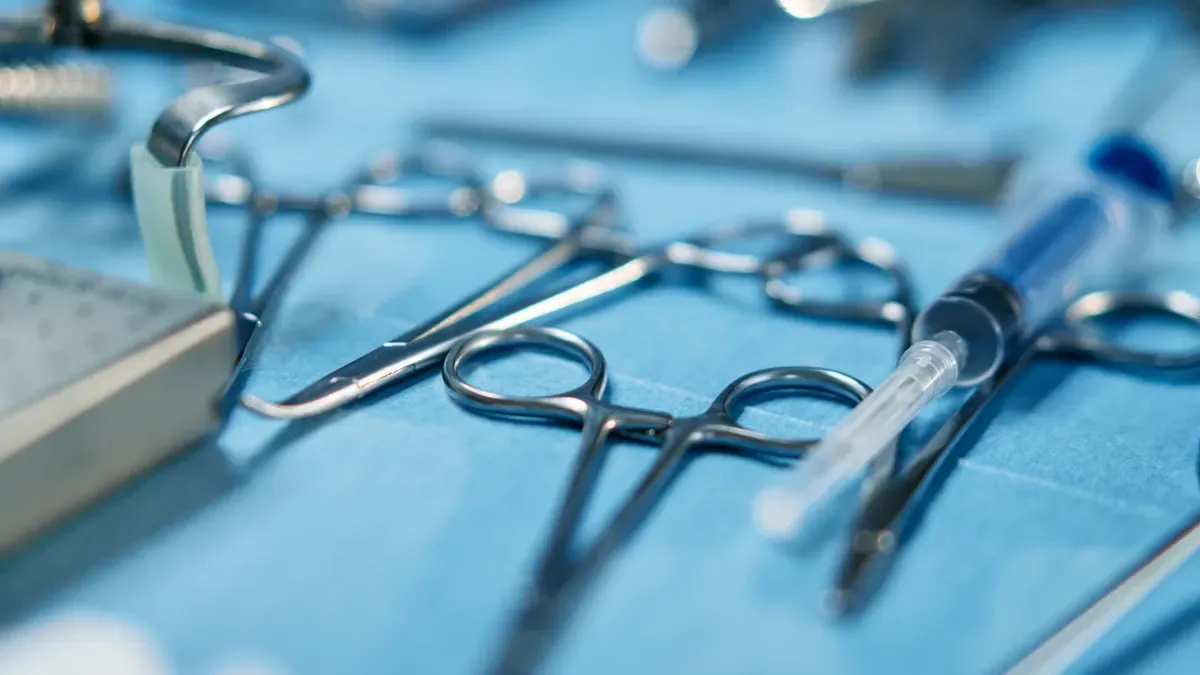
Graspers help doctors hold and move tissues. You use them to pick up or push organs. They look like long sticks with jaws at the end. Some jaws are smooth. Others have teeth or holes. You can pick the grasper you need for each job. Graspers are one of the Essential Instruments for safe surgery.
Function
Graspers do many jobs in surgery. They let you work with tissues without big cuts. Here are some ways you use graspers:
- Hold tissues so you can work carefully.
- Control blood vessels to stop bleeding.
- Help with suturing by holding tissues still.
- Pass other tools into the surgery area.
- Support organs during different steps.
Tip: Check your grasper’s jaws before surgery. Smooth jaws are good for gentle tissue.
Features
Different graspers help you handle tissues in special ways. Some protect soft tissues. Others grip tightly. The table shows how each type works and helps you avoid injury:
| Grasper Type | Function | Impact on Tissue Handling and Trauma Reduction |
|---|---|---|
| Dissecting Graspers | Used for careful tissue separation and blunt dissection. | Helps you find tissue planes and lowers injury. |
| Atraumatic Graspers | Have smooth or fine jaws for gentle handling. | Lowers injury and keeps tissue safe when you move it. |
| Traumatic Graspers | Have teeth or serrations for strong grip on thick tissue. | Hold tissue tight but lower the chance of damage. |
| Specialized Graspers | Have jaws made for certain body parts. | Give you more control and lower risk in special surgeries. |
| Fenestrated Graspers | Have holes in the jaws to change how they hold tissue. | Let tissue sit in the jaws and lower surface pressure. |
You can choose the best grasper for each part of surgery. This helps you protect tissues and feel confident.
Dissector
Description
A dissector helps you separate tissues in laparoscopic surgery. It looks like a long, thin stick with a special tip. The tip can be straight or curved. You choose the right dissector for each task. Some dissectors move tissues apart gently. Others cut through tough layers. You need dissectors to reach organs and blood vessels without making big cuts.
Here is a table that shows the main types of dissectors and what they do:
| Type of Dissector | Function | Role |
|---|---|---|
| General Dissectors | Used to separate tissue layers and planes | Important for isolating organs, blood vessels, and other parts during surgery. |
| Maryland Dissector | Has a slender, curved tip for many uses | Helps with sharp and blunt dissection, separates tissue planes, and exposes vessels and ducts. |
Function
Dissectors help you work safely and carefully. You use them to find the right tissue planes. They let you move tissues apart without hurting them. You can see blood vessels and ducts. This helps you know what to fix or remove. You use dissectors in many steps of surgery. They are one of the Essential Instruments that help you avoid mistakes.
Tip: Always check your dissector’s tip before surgery. A clean and sharp tip helps you work faster.
Features
Dissectors have special features that make your job easier:
- Articulation and flexibility help you move the tip in tight spaces.
- Enhanced visualization gives you a clear view of tissues.
- Atraumatic grasping keeps tissues safe from harm.
- Ergonomic handles make your hands comfortable during long surgeries.
- Electrocautery lets you cut and stop bleeding at the same time.
These features help you work with precision and keep your patient safe. Good dissectors help you finish surgery faster and lower the risk of problems.
Laparoscopic Scissors
Description
Laparoscopic scissors are used to cut tissues in surgery. You use them through small holes in the body. They have a long handle and a sharp blade at the end. You hold the handles outside the body to control them. There are different types for different jobs. Metzenbaum scissors have curved blades for gentle cutting. Mayo scissors have strong blades for thick tissues. The ring handles help you hold them tightly. The pivot part makes cutting easier. Stainless steel keeps the scissors strong and stops rust.
Note: Always check if the blade is sharp before surgery. A sharp blade helps you work quickly and safely.
Function
Doctors use laparoscopic scissors to cut tissues and blood vessels. They also cut sutures. These scissors let you make careful cuts without big openings. You can separate tissues gently and keep organs safe. They help you remove tissue and fix areas that need repair. You can trim mesh or other materials with them. Laparoscopic scissors are important tools that make surgery safer and faster.
Features
Laparoscopic scissors are better than regular scissors in many ways. The long handle lets you reach deep spots without big cuts. You get more control and can cut more carefully. The design helps lower injury and infection. Patients lose less blood and heal faster. The table below shows how laparoscopic scissors and regular scissors are different:
| Feature | Laparoscopic Scissors | Traditional Surgical Scissors |
|---|---|---|
| Design | Long handle for small openings | Short handle for direct use |
| Precision | More precise cutting | Regular cutting ability |
| Patient Trauma | Less injury from smaller cuts | More injury from bigger cuts |
| Infection Risk | Lower infection risk (4.76% vs 9.33%) | Higher infection risk |
| Blood Loss | Less blood lost (120mL vs 170mL) | More blood lost |
| Recovery Time | Faster healing because of small cuts | Slower healing time |
Tip: Pick the right scissors for each job. This helps you protect tissues and helps patients get better.
Energy Devices
Description
Energy devices help doctors cut, seal, and stop bleeding. These tools make surgery fast and safe. They use electricity, sound, or light to work. Each device has its own way of working. Some use strong electric currents. Others use sound waves or focused light. You see these devices in almost every operating room. They are important for minimally invasive surgery.
| Energy Device | Mechanism of Action | Applications |
|---|---|---|
| Monopolar Electrosurgery | Uses electric current to make heat for cutting and stopping bleeding. | Cuts tissues and controls bleeding. |
| Bipolar Electrosurgery | Electric current stays between two electrodes to keep heat in one place. | Stops bleeding and seals small blood vessels. |
| Ultrasonic Energy | Uses fast vibrations to cut and stop bleeding at the same time. | Used in gallbladder, appendix, and colon surgery. |
| Laser Energy | Uses focused light to burn or stop bleeding with care. | Used in gynecology and treating endometriosis. |
| Argon Beam Coagulation | Uses argon gas to carry energy for surface bleeding control. | Helps stop bleeding in liver or spleen surgery. |
Function
Doctors use energy devices to cut and seal tissues. They also stop bleeding and close blood vessels. These tools help avoid big cuts. You can stop bleeding fast, which keeps surgery safe. Energy devices help doctors work carefully and lose less blood. Newer devices make surgery faster and safer. Ultrasonic devices cause less heat damage, so tissues heal better. Laser energy in robotic surgery helps patients leave the hospital sooner and get hurt less.
Tip: Always check your energy device settings before surgery. Using it right keeps your patient safe.
Features
Energy devices have features that help doctors:
- You can change the power to fit the tissue.
- Alarms warn you if the device gets too hot or breaks.
- Handles are made to be easy to hold for a long time.
- Some devices can cut and stop bleeding at once.
- New designs keep heat from spreading to healthy tissue.
Doctors pick the best device for each job. This helps patients heal better and makes surgery go smoothly.
Needle Driver
Description
A needle driver helps you hold needles during surgery. It looks like a long clamp with jaws at the end. The jaws grab the needle tightly. This lets you push the needle through tissue. Most needle drivers have rounded edges. These edges protect the suture material. Some needle drivers have a knot pusher built in. You control the tool from outside the body. This makes it easy to use through small cuts. Needle drivers are one of the Essential Instruments for safe suturing.
Note: Always check the grip and jaws before surgery. A strong grip helps you avoid mistakes.
Function
Needle drivers help you close wounds inside the body. You use them to hold and move needles. This lets you stitch wounds or surgical cuts. The tool gives you a firm grip. You can push the needle through tissue accurately. Rounded jaws keep the suture safe. Many needle drivers have a knot pusher. This lets you move knots into place without hurting tissue. You can tie knots outside the body and push them in.
Here is a table showing what needle drivers do:
| Function Description | Purpose |
|---|---|
| Grasping and moving needles | Lets you stitch wounds or surgical cuts |
| Giving a strong grip on the needle | Helps you push the needle through tissue correctly |
| Rounded jaw edges | Keeps the suture from getting damaged |
| Built-in knot pusher | Lets you push knots into the body after tying them outside |
Tip: Practice using the needle driver with different needles. This helps you get better and faster.
Features
Modern needle drivers have features that help you work well. Automated needle grabbing lets you grab the needle again without extra steps. Built-in knot pushers make tying knots easier and more reliable. Ergonomic handles help your hands feel comfortable and let you stitch better.
Here is a table showing how features help you:
| Design Feature | Description | Benefit |
|---|---|---|
| Automated Needle Grabbing | Lets you grab the needle again easily | Saves time and makes work faster |
| Built-in Knot Pushers | Helps you tighten knots | Makes knot tying easier and more accurate |
| Ergonomic Handles | Handles are made for comfort and good grip | Helps you stitch well and keeps your hands safe |
You can pick the needle driver that fits your hand and needs. These features help you work quickly and keep your patient safe.
Surgical Mesh
Description
You use surgical mesh to support weak or damaged tissue during laparoscopic surgery. Mesh looks like a thin sheet or net. Doctors place it inside the body through small cuts. Mesh can be made from synthetic materials or processed animal tissue. Each type has its own purpose. You choose the mesh based on the patient’s needs and the type of repair.
| Type of Surgical Mesh | Material Type | Indications |
|---|---|---|
| Synthetic Mesh | Knitted or Non-knitted | Hernia repair, tissue support |
| Animal-derived Mesh | Processed animal tissue | Absorbable options for tissue repair |
Function
You use surgical mesh to fix hernias and support tissues. Mesh holds organs in place and helps prevent them from moving. Mesh gives strength to weak areas. You can use mesh for many types of repairs. Mesh helps tissues heal and lowers the chance of the problem coming back.
Note: Mesh is one of the Essential Instruments that helps you do safe and strong repairs.
Features
Mesh comes in different shapes and sizes. You can pick mesh that absorbs into the body or stays in place. Mesh can be soft or firm. Some mesh lets tissue grow through it. You must choose the right mesh to lower risks. Using synthetic mesh may cause chronic pain after surgery. Avoiding synthetic mesh can lower complications. When you respect natural anatomy, you help patients heal better.
- Mesh can be absorbable or permanent.
- Mesh may allow tissue to grow through it.
- Mesh can lower the chance of hernia coming back.
- Mesh choice affects pain and healing.
Studies show that laparoscopic repairs with mesh have different complication rates compared to open surgery. You must pick the best mesh for each patient to help them recover well.
You need Essential Instruments to do laparoscopic surgery safely. Each tool helps you work carefully and lowers the chance of hurting someone. Training and practice help you use these tools quicker and better. New technology, like robots and smart devices, keeps making surgery safer and helps patients get better results.
FAQ
What is the main benefit of laparoscopic surgery?
Doctors make smaller cuts, so you heal faster. You have less pain and fewer scars after surgery.
How do you clean laparoscopic instruments?
You need to wash, disinfect, and sterilize each tool.
| Step | Action |
|---|---|
| Wash | Take off dirt |
| Disinfect | Kill germs |
| Sterilize | Use heat |
Can you reuse laparoscopic instruments?
Some metal tools can be used again after sterilizing. You must throw away plastic tools that are for one use only.